
Small white dots on fingernails are usually punctate leukonychia from minor matrix trauma, not a zinc emergency. Grossman, Scher, Zaias, and Baran mapped what the spots mean — and when a nail change is not ordinary.
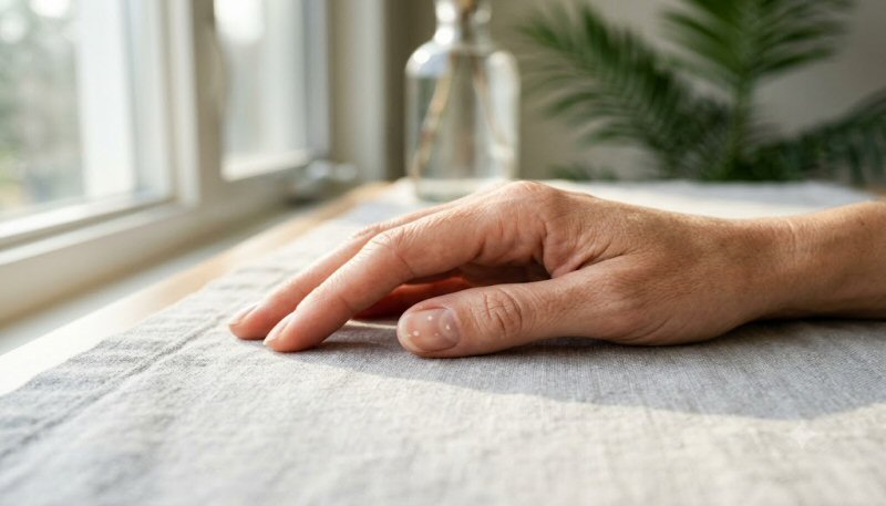
You notice them while washing a mug or after a manicure comes off: one or two chalky dots on a fingernail, sometimes a scatter of them, sometimes a thin white line. The internet offers a confident diagnosis before you finish the search. Zinc. Calcium. A liver problem. A hidden illness that has finally written itself on the nail.
Most of the time the nail is not writing a mineral report. It is writing a small accident that happened weeks ago, under the proximal fold, where the matrix was making the plate. A knock against a counter, a too-aggressive cuticle push, a child who slammed a door on a fingertip — any of these can leave a white island that only becomes visible once that segment of nail has grown out far enough to see.
Dermatologists call the common version leukonychia punctata: true white spots inside the plate, not surface chalk you can wipe away. Understanding the difference between a matrix bruise, a surface stain, and a rare systemic pattern is what turns a scary photograph into a useful observation.
What the White Spot Actually Is
A normal nail plate is a sheet of compacted keratin made by the matrix under the proximal nail fold. When that sheet is compact and aligned, light passes through and reflects off the pink nail bed beneath. You see a translucent plate and a pink bed. When a patch of matrix is briefly injured, the keratin in that patch is laid down with more air spaces and less orderly packing. Light scatters. That scatter looks white.
Nardo Zaias, whose work on nail growth and pathology still anchors the field, described how matrix insults travel forward with the plate like a time-stamped mark. The spot does not sit on the surface. It is in the plate. You cannot scrape it off. It will reach the free edge in weeks to months and then be cut away.
Two other white appearances get mixed in and should be separated:
- Apparent leukonychia — the plate is normal; the bed looks pale because of pressure, anemia, or edema. Press the nail and the color may change.
- Pseudoleukonychia — surface change such as white superficial onychomycosis or keratin debris. The white can often be scraped or is limited to the outer layer.
The ordinary dots people photograph after a manicure or a clumsy week are true punctate leukonychia. They are opaque, well-defined, and move distally as the nail grows.
Why Trauma Is the Usual Author
The matrix is a thin germinal strip hidden under the cuticle. It has no padding. A modest knock that you forgot by dinner can still disorder a few columns of keratinocytes. Manicure trauma is a classic source: pushing the cuticle hard, filing the plate too thin, or using a metal tool under the free edge that leverages force back toward the matrix. Children get spots after playground knocks. Keyboard-and-phone hands get them from repeated small hits against desks and device edges.
Philip Fleckman and others who mapped nail-unit anatomy emphasized how little force it takes to disturb matrix output without breaking the skin. That is why the history is often blank. People say, “I didn’t injure it.” They mean they did not see blood or a bruise. The matrix does not require a visible bruise to misfire for a day.
Fingernail growth is roughly 3 millimeters a month in adults, slower in toes and in older age. A spot that appeared “suddenly” near the lunula was made about a month earlier. A spot near the tip was made two or three months earlier. The calendar is useful. If new spots keep appearing at the lunula every few weeks, something is still hitting or inflaming the matrix. If old spots only march forward and no new ones form, the insult is over.
The Zinc Myth — and What Nutrition Actually Does
White spots became a folk marker for zinc deficiency because severe zinc deficiency can disturb keratinization. That part is not invented. What is invented is the leap from two dots on an index finger to a diagnosis of mineral collapse.
True acrodermatitis enteropathica and severe acquired zinc deficiency produce more than scattered nail dots. They produce rash around the mouth and extremities, diarrhea, hair loss, and often a more diffuse nail dystrophy — not a single tidy fleck. Calcium folklore is even thinner. The plate is not a calcium warehouse. Dietary calcium does not print white islands.
When nutrition does mark nails, the pattern is usually different. Beau’s lines are transverse grooves after a severe illness or chemotherapy pause in matrix output. Muehrcke’s lines are paired white bands that do not move with the plate and point to apparent leukonychia from hypoalbuminemia. Terry’s nails and half-and-half nails change the whole bed color. Those are systemic patterns. They do not look like a few random dots.
If diet is poor, the honest work is still food quality, not treating a fleck as a lab test. A spot is a weak reason to start high-dose zinc. Excess zinc can itself disturb copper balance.
Other Common, Still-Benign Patterns
Transverse white lines after a fever or a slammed door. A single horizontal band across one nail often dates a matrix shock. If every nail shows a line at the same height, the shock was systemic — high fever, severe illness, or a drug pause — and the lines are cousins of Beau’s lines.
Habit-tic and cuticle picking. Repeated pushing or picking at the proximal fold can leave a washboard nail and scattered white marks on the thumbs. The habit is the treatment target, not a supplement.
Occupational microtrauma. Chefs, mechanics, string musicians, and people who type with long nails all feed the matrix small insults. The spots cluster on the working fingers.
After acrylics or harsh acetone weeks. The plate can look chalky from dehydration and layering, which is closer to surface change. True matrix spots can still appear if the cuticle was forced back for weeks.
When White Nails Are Not Ordinary Dots
Most photographs of “white spots on nails” are punctate leukonychia. A smaller set deserves a closer look:
- White that spreads as a crumbling, chalky patch from the free edge — think superficial white onychomycosis, which Nardo Zaias described as a surface fungal pattern, often Trichophyton mentagrophytes.
- A nail that turns white, lifts, and thickens — onycholysis plus dystrophy, not a spot.
- Total leukonychia of all nails from childhood — a rare inherited keratin defect, not a new deficiency.
- Paired white bands that do not grow out (Muehrcke) plus swelling or known liver or kidney disease.
- New pigment, a streak that reaches the fold, pain, or a nail that stops growing — those are different problems and do not belong in the “dots” conversation.
Robert Baran’s clinical nail texts and the reviews by Richard Scher and Marc Grossman keep repeating the same triage: pattern, number of nails, whether the white moves with growth, and whether the rest of the skin and health story is quiet.
Hidden Triggers People Miss
The insult is often not the dramatic door slam. It is the weekly manicure that treats the cuticle as an obstacle. It is using the nail as a tool to open packages. It is a tight glove or a sport that jams fingertips. It is a toddler who grabs fingers. It is onychophagia — nail biting — which both injures the matrix and invites surface infection.
Seasonal dryness can make spots more visible without creating more of them. A dehydrated plate scatters more light. The same person who barely noticed dots in humid summer sees them clearly in heated winter air. That is optics plus a drier plate, not a new disease.
Medication and chemo can pause or disorder matrix output across many nails at once. Those marks are useful dates of treatment, not random freckles.
Myths vs Facts
Myth: White spots mean you need zinc or calcium.
Fact: Isolated punctate spots almost never diagnose a mineral deficit. Severe deficiency looks like a systemic skin and gut story, not two flecks.
Myth: You can rub the spot off if you file enough.
Fact: True leukonychia is inside the plate. Filing only thins a healthy nail. Surface chalk or fungus is the exception, and even then filing is not a treatment plan.
Myth: A white spot is an infection starting.
Fact: Ordinary dots are packed keratin with air. Infection usually changes texture, smell, or the fold, or it advances as a crumbling white patch from the tip.
Myth: Darker nail beds hide problems; pale spots are always serious.
Fact: Contrast makes spots easier to see on some plates. Visibility is not severity.
Myth: If a child has spots, their diet has failed.
Fact: Children hit their hands constantly. Punctate leukonychia is one of the most common pediatric nail findings and usually graduates off the free edge without a supplement.
How to Manage Ordinary White Spots
You do not treat the spot. You stop making new ones and wait for growth.
Leave the cuticle alone. It is a seal, not a rim to be carved. If you want tidy folds, soften with a bland oil and push gently after a shower — do not scrape the matrix neighborhood with metal.
Stop using nails as tools. A key, a letter opener, or the side of a finger saves the plate.
Keep the plate slightly flexible. Very dry nails crack and invite more matrix stress at the fold. A plain emollient on the folds at night is enough for most people. You do not need a miracle keratin serum.
If you wear enhancements, give the matrix recovery time between sets. Forcing a lift or prying off acrylics is a reliable way to print a row of future spots.
Photograph one nail next to a ruler once a month if you are the worrying type. Watching the spot march forward and disappear is more calming than another search.
Diet still matters for the whole integument — protein, vitamin C for collagen cross-links, and enough zinc from food if intake is truly low — but that is general nail and skin maintenance, the same logic behind why collagen still expects load and vitamin C. It is not a prescription written by two dots.
Hair and nails share keratin biology and a long memory for stress and season. A person who also notices extra shed in spring or after illness is often seeing the same system on a different clock, which is why hair and nails still reflect seasonal biology belongs in the same mental folder — not because spots are seasonal, but because keratin appendages keep a delayed diary.
When to See a Doctor
Book a clinician — dermatology if you can — when any of these are true:
- White change is crumbling, spreading from the tip, or paired with thickening, lift, or fold swelling.
- Every nail suddenly shows bands or complete whitening.
- Spots come with rash, hair loss, diarrhea, or known malabsorption.
- A dark streak, bleeding under the nail without injury, or pain that does not match a remembered knock.
- A child has nail destruction, not just dots.
A single fleck that grows out is not an emergency. A changing nail unit is a different appointment.
FAQs
Do white spots mean I am low in zinc?
Almost never if the only finding is a few punctate marks. Zinc deficiency severe enough to change nails usually brings rash, gut symptoms, and more diffuse dystrophy. Food-first intake is reasonable; megadose zinc because of a fleck is not.
How long until a spot grows out?
Fingernails advance about 3 mm per month. A mark near the lunula can take three to five months to reach the free edge. Toenails are slower, often twelve to eighteen months for a full length.
Can a manicure cause them?
Yes. Aggressive cuticle work is one of the most common adult causes. The spot appears later, so the manicure and the mark do not feel connected unless you line them up on a calendar.
Should I take calcium for white nails?
No. The plate is keratin, not a calcium crystal. Calcium folklore is a category error.
Are white spots in children a sign of poor diet?
Usually no. Playground and door-frame trauma explain most pediatric dots. Look at growth, energy, skin, and diet as a whole, not at one thumbnail.
Can fungus look like white spots?
Superficial white onychomycosis can look chalky, often as a patch rather than a neat embedded dot, and it may be scrapable. If the white is soft, spreading, or limited to the surface, a clinician can confirm with a sample rather than a guess.
Conclusion
A white spot on a nail is usually a delayed photograph of a small matrix injury. It is not a lab printout of zinc or calcium. It cannot be scrubbed off because it is built into the plate. It will walk off the end of the finger if you stop repeating the insult.
Save the worry for patterns that do not behave like a traveling fleck: crumbling fungus, bands across every nail, lift and pain, or a body that is clearly unwell. For the ordinary dots, the useful moves are gentle folds, fewer tool-nails, and patience measured in millimeters per month. The plate is honest. It just keeps a slower clock than the search bar.